
By : Urvinder Kaur, Alex Muller, Lauren Harnois, and Anna Kuruc
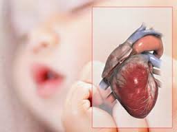
It is very important to note that congenital heart defects are among the most common birth defects affecting more than 40,000 children a year. The defects can be very complex and life threatening or relatively benign. Treatment is different for every cardiac defect and usually involves medicines, surgeries, heart transplants, and mechanical ventilation.
Hypoplastic left heart syndrome (HLHS) is characterized by hypoplasia of the left ventricle, aortic stenosis, hypoplasia of the ascending aorta, and coarctation (narrowing) of the aorta. In normal conditions the left side of the heart receives oxygenated blood from the lungs and pumps it into the systemic circulation. In patients with HLHS, the left ventricle and the aorta are underdeveloped. The blood from the lungs goes to the right heart via an atrial septal defect and is pumped to the rest of the body by the right ventricle through the patent ductus arteriosus (PDA). This defect is a ductal dependent syndrome, and can be catastrophic if the PDA closes. Babies are often cyanotic at birth to varying degrees, depending on the severity of their defects. The quick and life saving diagnosis is made by fetal ultrasound during pregnancy. HLHS is often present in kids who have other genetic disorders such as Trisomy 13, 18, etc. Maternal obesity affects the normal development of the fetal heart. A study found a direct correlation between increasing BMI and increased cases of congenital heart defects such as HLHS.1
Initial treatment for HLHS includes stabilization with prostaglandins for ductal patency and initiation of pulmonary hypertension by methods such as hypoventilation and using sub-ambient air to maintain systemic perfusion. Surgical palliation treatment includes the Norwood procedure, followed by Glenn and finally the Fontan procedure. Prognosis has improved with the introduction of the Norwood procedure. Today, 70% of HLHS patient have reached adulthood.
Another important shunt is the atrial septal defect (ASD). Many neonates live well into adulthood without a clinical diagnosis. ASD is a congenital defect where the interatrial septum is missing and oxygenated blood from the left side mixes with the deoxygenated blood from the right side of the heart. Diagnosis is made through ultrasound, echocardiogram, EKG, and physical exam. Mothers who smoke during pregnancy can increase their chances of having a baby with ASD and pulmonary valve anomalies.2 Treatment of a small asymptomatic ASD does not require therapy, while an ASD greater than 9mm needs surgical intervention. Options include direct suture anastomosis or patch closure and autologus pericardium or synthetic material. More recently, a transcatheter device has become the treatment of choice. Infants have a better prognosis when defects are corrected before signs of pulmonary hypertension develop.
The last cardiac defect that we would like to mention is the tetralogy of fallot (TOF). TOF involves four anatomical abnormalities of the heart: a large VSD, a stenotic pulmonary artery, an overriding aorta, and right ventricular hypertrophy. The degree of cyanosis of TOF depends largely on the size of the right-to-left shunt. Patients with left-to-right shunting may have little to no cyanosis and are referred to as “pink tetralogy.” Patients with TOF may experience episodes of extreme cyanosis called “tet spells” often during times of stress such as crying, dehydration, and fever. If left untreated these hypercyanotic episodes can be fatal. Deletion of the gene on Chromosome 22, (22q11) is often the cause of TOF among other congenital heart defects. Another gene associated with this anomaly is the jagged1 gene, which can be diagnosed through the images of an echocardiogram. On an anterior-posterior chest x-ray, the heart is shaped “boot-like.” 3
Treatment after birth includes administering prostaglandin E to maintain the ductal patency and pulmonary perfusion. In some cases Blalock-Taussig shunt (BT shunt) can be performed to provide adequate pulmonary blood flow to the lung. Surgeries are done over time from neonates to 4-6 months of age. Mortality depends on the severity of TOF. It carries a 35% mortality rate if left untreated in the first year of life and 50% mortality rate in the first three years of life. Genetic testing to look for these mutations can be a viable option to couples planning a pregnancy.4
In conclusion, respiratory therapists play a key role in caring for infants with cardiac defects, including initiation and management of mechanical ventilation post-operatively. It is essential to monitor hemodynamics to implement the correct therapy. An arterial line provides a continuous blood pressure monitoring as surrogates of end-organ blood flow. Noninvasive monitors such as near-infrared spectroscopy measure venous oxygen saturation in different parts of the infant’s body to follow different cardiac trends. It is also important to monitor serum lactate, neurological status, and urine output.3
References
- Madsen, Nicolas L., Stephen M. Schwartz, Mark B. Lewin, and Beth A. Mueller. “Prepregnancy Body Mass Index and Congenital Heart Defects among Offspring: A Population-based Study.” Congenital Heart Disease 8.2 (2012): 131-41.
- Sullivan, Patrick M., Leslie A. Dervan, Sheridan Reiger, Sujatha Buddhe, and Stephen M. Schwartz. “Risk of Congenital Heart Defects in the Offspring of Smoking Mothers: A Population-Based Study.” The Journal of Pediatrics 166.4 (2015)
- Walsh, Brian K. Neonatal and Pediatric Respiratory Care. 4th ed. N.p.: Elsevier, 2015.
- Eldadah, Z. A. “Familial Tetralogy of Fallot Caused by Mutation in the Jagged1 Gene.” Human Molecular Genetics 10.2 (2001): 163-69.

Anna and team very interesting blog about congenital heart defects. Currently, congenital heart defects are the most common birth defects affecting large numbers of infants in US. Furthermore, respiratory therapists are highly involved in caring for infants who are critically ill due to cardiac defects. I totally agree with your analysis of the types of congenital heart defects that you and your teams focused on. Indeed, respiratory therapists are involved in the initiation and management of mechanical ventilation post operatively, so that being familiar with the pathophysiology and clinical manifestations of those defects would help RTs provide good care.
LikeLike
Thank you for sharing this interesting blog, “Tiny Hearts, Enormous Defects,” discussing congenital cardiac defects because it impacts the lives of more than 40,000 children every year in the United States alone. The severity and complexity of treatment vary based on the location and size of the cardiac defect. That is why it is so essential for respiratory therapists to quickly assess and recognize the clinical manifestations for the different cardiac defects, in order to initiate appropriate treatment in a timely manner. Tetralogy of fallot (TOF) often manifests with extreme periods of cyanosis, prolonged crying and syncope. Treatment for this type of cardiac defect would focus on maintaining pulmonary perfusion through the ductus arteriosus because of the shunt present between the right and left ventricles as well as the narrowing of the pulmonic valve, causing the aorta to override blood flow. Another type of ductal dependent cardiac defect is the hypoplastic left heart syndrome (HLHS). This is a ductal dependent shunt because oxygenated blood would not be able to systemically circulate since the left side of the heart is underdeveloped and not able to function properly. Normally this shunt between the pulmonary artery and aorta would close shortly after the infant is born due to the increase in PaO2 levels with a decrease in PaCO2 levels. However, prognosis is poor for TOF and HLHS if the shunts are closed prior to surgical intervention. Not all cardiac defects are ductal dependent, such as the atrial septal defect (ASD). Only symptomatic shunts greater than 9mm may require surgical repair, by closing the opening between the right and left atrium.
LikeLike